
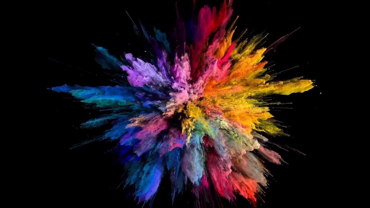
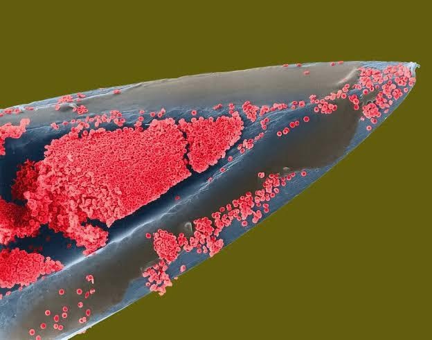
The colors are added in, of course, with it being an electron microscope image. Another picture:
Its crazy how crude all of our tools look at this magnification.
Some medical tools look crude even at regular size… they don’t call orthopedics bone carpenters for nothing!
People would never set foot in a hospital again if they found out how many orthopedic surgeries involve a dewalt drill at some point.
My knee replacement was carried out with an epidural pain block, plus sedation. I came down from cloud nine briefly to wonder why someone was doing renovations while surgery was in progress - then realised all the drilling and hammering was my new joint going in. Phew! Back to lala land…
Lmao “oh shit I’m a house”
I’m just going to leave this discovery here.
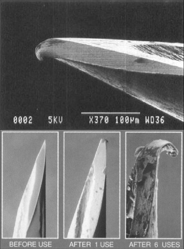
That’s great. I rotated through an ortho lab in the 1990s, and the joint replacement kits back then included a sterile, disposable drill that you were just supposed to throw out after the procedure.
I recently saw a knee replacement that used one of those ryobi oscillating cutters (the ones that were super trendy a few years back). Total garbage for home use, but man with a 3D printed cutting guide shaped to fit over the bone, they finished the osteo and arthroplasty portions in ten minutes flat. Just insane what we can accomplish when we combine modern volumetric imaging techniques with coupons for home depot.
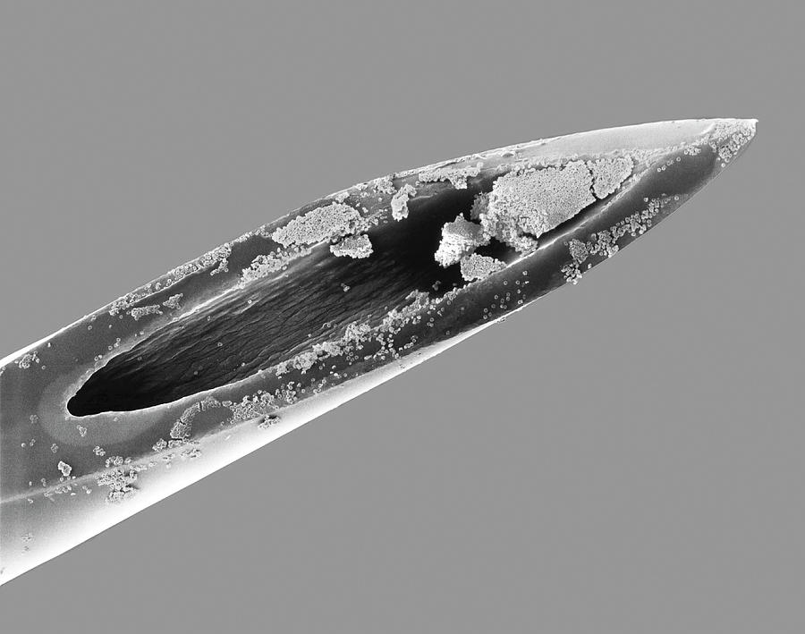
I gained an appreciation for how precise/sharp our tools are when I learned microtomy. If you so much as touch the cutting edge with anything outside of its intended use it messes up that area of the blade instantly. Same goes for a nice pair of chef’s knives.
It actually looks a LOT smoother and sharper than I expected. Look at microscope photos of razors and knives and they look like chewed up chisels.
See that little hook at the point? This is from penetrating skin ONCE.
This is why you don’t re-use needles folks!
There are other reasons.
I can’t believe it’s that fine or how easily human skin can bend it. I guess our skin is a better protector than I’d given credit.
All these images are super interesting and I’m truly glad they got posted.
Holy shit. I noticed that too and thought that must be one of the ones that hurts going in, figured it was from when they draw the medicine into the syringe, or maybe even from taking the cap off.
Crude aspects of fleshy meatbags.
From the moment I understood the weakness of my flesh, it disgusted me. I crave the certainty of steel.
certainty of steel.
Yeah… take a few material courses in engineering…
It’s not so certain a lot of the time…
I crave the certainty of neutronium
I want to eat a red blood cell. Like one the size of my hand that tastes like a gummy bear
Can’t you just eat some real gummy bears? I think they even make big ones.
Frighteningly big.

Please do not buy your child a gummy bear bigger than their head. We have enough problems with diabetes as it is.
Or use your own blood to make gummy bears from it.
Or buy regular gummy bears, put in a bath of your blood and let them swell up!
This is fascinating. I mean we all know the theory, but to actually see the cells under magnification puts you in range, and makes you wonder what else there is to know. And the answer is always MORE.
Education should work more practical application in with the theory. I’m looking at you, calculus!
Seriously. I’m in my 40s and this is the first time I’ve ever had any sense of scale for red blood cells. Very cool!
Where is the plasma?
I’m assuming if the syringe was wet before being placed in the microscope, the vacuum of the chamber would cause most of the water in the plasma to vaporize. The remaining salts and compounds would be much smaller than the red blood cells. The density of the red blood cells would be much larger than any remaining plasma, so the bulk of your backscattered electrons will be coming from the cells and needle, making the plasma essentially transparent. This is a fairly low magnification image for SEM, but that’s how you get such fantastic depth of field.
how were the colours added? like do you carefully select each isolated cell to add the colour or is there some kind of algorithm?
When I segmented 3D MRI and CT scan images before I used the contrast borders for help a lot. There were some algorithms for finding edges that you could tune by setting search radiuses and thresholds. There was also an option of growing an area by a certain amount of pixels outward, and then threshholding the result back down to only the brighter parts, that kind of thing. You had to be a little clever about how you’d combine it. And ultimately, sometimes I just had to add and subtract a few points manually.
Segmenting is more assigning areas to distinct objects (separating bones from the rest in my case), but you could totally use it as a basis for coloring, so I assume the process is similar here.


